What is Dermatofibroma?
Dermatofibroma also known as histiocytomas are noncancerous skin growths which are quite common. They are firm to hard and can be tender. These bumps or growths normally continue for life and may heal to become depressed like scars after several years. Sometimes, dermatofibromas found grouped in large numbers of linear clusters are seen linked with immune disturbances such as HIV, leukemia as well as lupus.
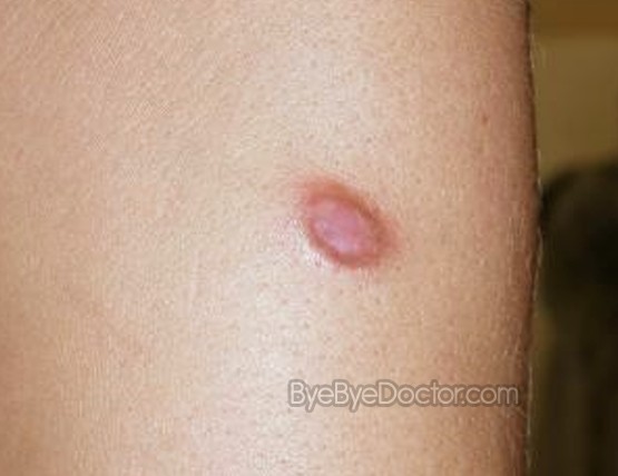
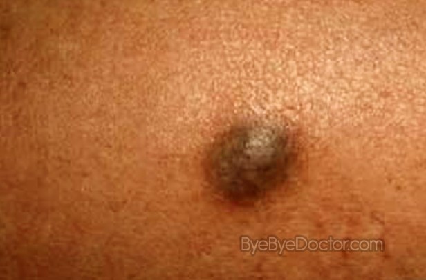
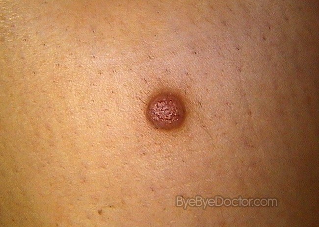
Dermatofibromas are usually small and pea size and occur either on the skin of the legs or the arms. They can range in color from yellow-brown to dark red or reddish brown. They are composed of spindle cells or histiocytes in association with collagen.
Dermatofibroma Causes
It is not certain what the exact cause of dermatofibroma is mostly because of its nature of being persistent. The possible causes are as:
Injury
An injury to the skin which is minor such as a prick from a thorn and can lead to dermatofibroma.
Insect bite
It can even be caused by a bite of an insect.
Sex
Women are more likely to have this condition than men.
Age
Usually develop in middle-aged adults and rarely found in children.
Heredity
Dermatofibroma may also be caused by family history although this is debated in the medical community.
Dermatofibroma Symptoms
Symptoms of dermatofibroma which are important include:
http://www.Symptoms-Causes-treatment.blogspot.com detect diseases at an early stage symptoms, and find out the causes and treatments best suited.
- Usually found on the lower legs but can be on the arms or trunk
- Raised from the skin and bleed if damaged
- Size vary – small as a BB pellet but as large as a fingernail
- Dimple inward when pinched
- Darker for individuals with dark skin
- Color may change over time
- The pink, red, gray, brown or purplish patches can be seen on the affected area of skin
- Usually painless but can be tender, itchy or painful
- Develop gradually normally over several months
- Be 5 mm to 10 mm across, rarely any bigger
- Have a shiny, dull or scaly surface
If you develop any type of lump your doctor will want to examine it. He or she will make the diagnosis by sight as well as touch. The doctor will also squeeze the skin over the lump – when squeezed together, a dimple will form. If there is any doubt about the bump, it can be surgically removed. The removed tissues are then examined by microscope.
If you have any type of new skin growth, it is wise to let your physician make an accurate diagnosis especially if you have any lesion which either changes color or is dark black or brown. If the growth bleeds, becomes painful or grows quickly then you need to see your physician immediately.
Dermatofibroma Treatment and Removal
Dermatofibroma are really best ignored. But if diagnosis is uncertain, a piece needs to be removed for analysis of the tissue.
Treatment of dermatofibromas can be considered if they get in the way becoming irritated by clothing or get in the way of shaving.
There are two main methods of removing a dermatofibroma:
Since a dermatofibroma grows so deeply, total removal surgically requires cutting it below the surface level of the skin. This method normally leaves a very noticeable scar. Alternatively, the nodule can be flattened to the surface of the skin by shaving the top with a surgical knife or by freezing with liquid nitrogen. With both treatments, the top layers are destroyed but the layers which are deeper remain. The nodule can grow back after several years. If the lesions grows back or bleeds, or if there is any fear that it could be a skin cancer, the growth needs to be biopsied or removed again.
Since these lesions are not dangerous and is not a condition which is painful, it is best to leave the bump alone. A hot damp cloth may be place over the bump to encourage some healing, but this is also underneath deliberation as to its usefulness. Bumps are not often medically treated unless the bump is in a bad location cosmetically or if the bump creates problems.
With the growth on the market of “Medical Tourism” hospitals, clinics as well as medical centers around the world offer Dermatofibroma Removal. The procedure is offered in India at the Moolchand Medcity where there are 400 doctors who are JCI accredited and ISO certified. This hospital treats over 7000 international patients each year as Medical Tourism becomes big business.
Dermatofibroma Pictures


