Monday, August 4, 2014

Shingles Rash

Shingles Rash
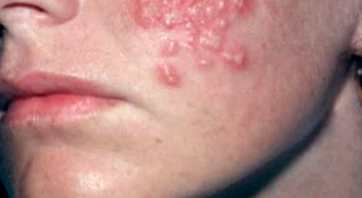
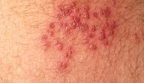
Shingles is a skin rash caused by varicella-zoster virus, a type of herpes virus, which causes a disease known as chickenpox. Shingles is an infection that occurs on the nerve and area of skin, which is supplied with nerve endings. People who have had chickenpox previously can develop shingles rash. Even those who have had mild chickenpox can also suffer from shingles. 


Herpes zoster virus is different from herpes simplex because the latter causes genital herpes, which is a different condition from shingles. It is estimated that 1 in 5 people are likely to develop shingles in their life. This condition can occur at any age but it is more susceptible to people over the age of 50. Rarely does shingles rash manifest more than once but about 1 in 50 persons are likely to develop shingles more than once. 


Causes


Mainly shingles rash may occur for no obvious reason but at other times, the condition may be caused by stress and illness triggers. The weakened and ageing immune system in older people may be one reason why they develop the rash. The immune system suppresses the virus and keeps it inactive thus preventing it from multiplying. Therefore, if there is slight weakened immune system, as it happens in older people, the virus may reactivate and multiply leading to development of shingles. 


Many people suffer from chickenpox at their childhood stage. When a child or a person suffers from chickenpox, the virus that causes this condition does not completely disappear and it remains dormant in body. Some virus particles remain in the nerve roots surrounding the spinal cord in an inactive state. When these particles are present in nerve roots, they do not cause harm there since they are inactive. They do not show symptoms. 


However, for reasons not clearly understood, these inactive virus particles may start multiplying or reactivating after some years of dormancy. When the virus is reactivated at the spinal cord nerve area, it travels along the nerve to the reach the skin where it causes shingles. Shingles is more prevalent in people with immunosuppressant or poor immune system. 


Young people with HIV and AIDS may suffer the condition. Similarly, people who have suppressed immune system as a result of use of treatment options like chemotherapy and steroids may also suffer from the condition. Being exposed to shingles may not cause the rash but if you get in contact with a person with shingles and you have not suffered from chickenpox before or you have not received chickenpox vaccine, there is possibility of suffering from chickenpox. 


You can be exposed to chickenpox if you come in contact with fluid from the shingles blisters.


Being over 50 years places you in a high risk of suffering from shingles. If you have recently had a surgery or you have been using cancer treatment medication like chemotherapy, then you may be susceptible to suffering shingles. Other risk factors include radiation therapy, AIDs, cancer, poor nutrition, stress, organ transplant, diabetes, and lupus, which suppress the immune system.  


Symptoms of shingles rash


The varicella-zoster virus mainly affects one nerve and on one side of the body. The skin area supplied with nerves begins showing symptoms like pain and rash. At times, two or more nerves that are close to one another are affected by the virus. The mainly nerves affected are those which reach the skin on abdomen, chest, upper face, and the eyes. 


Early symptoms of the condition include sensitivity to light, headache, and flu-like symptoms with no fever. The itching and tingling begin on the band of skin. A person with the condition has a localized band of pain. The pain can occur anywhere and it depends on which nerve is affected. The pain also ranges from moderate to severe. The pain may be constantly dull or at times, it may be gnawing pain. At other times, you may experience sharp and stabbing pain, which comes and goes. 


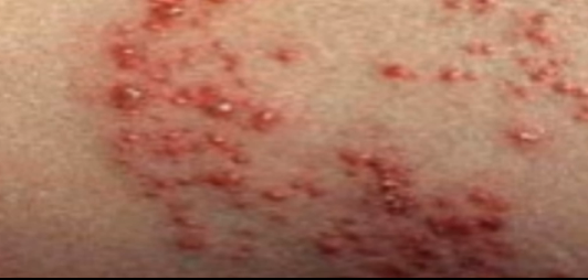
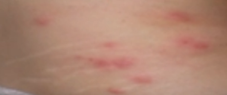
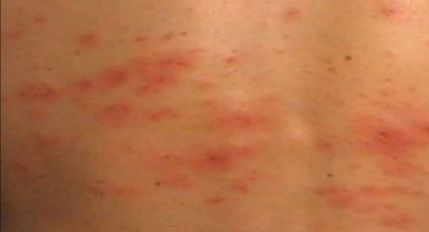
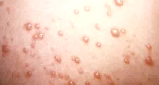
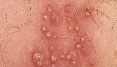
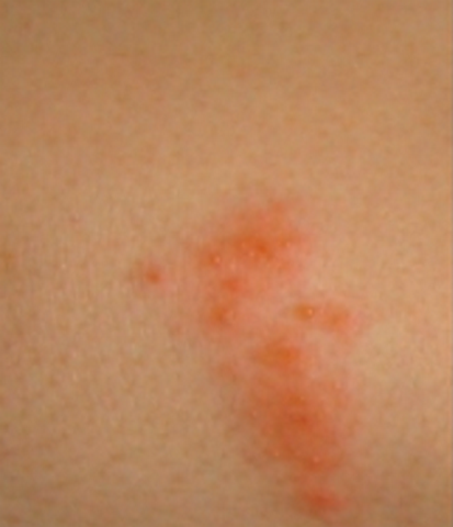
The affected skin becomes tender. A rash occurs within 2 to 3 days following the start of pain. First, red blotches appear and they develop into itching blisters. The rash appears like chickenpox but it only affects the area of the band of skin, which hosts the affected nerves. The tissue under and around the rash may become inflamed due to effects of the virus attack. The blisters dry and form scabs before they eventually fade away. The area where the blisters occurred may have slight scarring. 


Treatment


There is no cure for this skin condition but there are treatments, which can shorten the illness and prevent other health complications from occurring. Antiviral medicines are introduced to help relieve pain and shorten the duration of the shingles rash. To relieve pain, antidepressants and topical creams are applied. When treatment with antiviral is given in advance, it can help prevent later complications like postherpetic neuralgia. Antibiotics are used to stop infection of blisters. 


Shingles Rash Pictures


Lymphedema

Lymphedema
Lymphedema is a condition that causes swelling of one leg or arm but it can also at times cause the swelling of both legs or arms. The condition occurs when there is blockage of lymphatic system, which plays a role in circulatory and immune function. The swelling or inflammation is caused by build-up of fluid due to blockage on the lymphatic system. The lymphatic system consists of vessels that run throughout the body.


These lymph vessels collect excessive fluids, lipids, proteins and wastes from body tissue. The fluid is transported to lymph nodes where the waste is filtered and the fluid is returned to bloodstream. Primary lymphedema is an inherited condition that occurs due to anatomical abnormality within the lymph vessels. Secondary lymphedema occurs when there is damage or obstruction on otherwise previously normal lymph system.


Causes


The lymph system helps keep the body healthy by circulating fluid that collects microbes and waste products from the tissues and filtered by the lymph nodes. The wastes are filtered by lymphocytes, which are infection-fighting cells. After the filtering, the waste is flushed from body. When the lymph vessels cannot effectively drain the lymph fluid from an arm or leg, such a situation may lead to lymphedema.


Primary lymphedema is rare but it is an inherited condition. The cause of this condition may be due to Milroy’s disease, which starts at infancy and causes abnormalities on your lymph nodes. Another cause of primary lymphedema may be Meige’s disease, a hereditary condition that causes the lymph vessels to develop without valves that help regulate the backflow of the fluid. Lymphedema may also occur late at the age of around 35 years. This is called late onset lymphedema and is a primary form of the condition, which occurs due to abnormalities.


Secondary lymphedema is the most common form of this condition and is caused by a disease or condition that leads to the blockage of the lymph vessels. Damage of lymph vessels can be caused by a surgery where the vessels are cut such as during breast cancer surgery in which one or more nodes are removed from the armpit. If the retained lymph nodes and vessels cannot circulate the fluid as required, this may result to lymphedema.


Infection of the lymph nodes can cause restricted flow of lymph fluid. The blockage may also be caused by parasites that invade the lymph system. Filariasis causes blockage on lymph vessels where infestations by parasite called Wuchereria bancrofti occur in the lymph system. Cancer treatment methods like radiation and may also block the vessels or impair the function of the lymph nodes. The radiation can cause inflammation and scarring, which restricts the flow of fluid through the lymph system. A tumour growing near the lymph nodes could enlarge and cause blockage on the vessels making the flow of lymph fluid ineffective.


Symptoms of lymphedema


Lymphedema mainly affects one arm or leg but in rare cases, it can affect both arms or legs. The common symptoms of this condition include swelling of part of the arm or leg. At times, the swelling may cover the entire arm or leg including the toes and fingers. A person with the condition feels heaviness or tightness of the affected extremity whether the leg or arm. There is restricted range of motion accompanied by aching and discomfort in the extremity part of the body affected.


The affected limb may experience recurring infections. Other symptoms include hardening and thickening of skin on extremity. The swelling varies from mild to serious and while the mild swelling may have hardly noticeable change in arm size, on the other hand, severe swelling can occur which makes the limb impossible to move. If the lymphedema is caused by cancer, it may take months or several years before you notice the swelling.


The long-term accumulation of lymph fluid causes inflammation and scarred tissue, something that causes a firm and stiff swell, which may not retain its displacement when it is indented with a fingertip. The skin around the infected area may thicken and become lumpy, scaly, and cracked. The affected area also becomes tender, sore, and there is loss of mobility. Other symptoms include fatigue, weight loss, and embarrassment.


Treatment


Although there is no cure for this condition, it can be managed by taking care of the affected extremity. The treatment therapies help in reducing inflammation and pain. Light exercise of the limb enhances the movement of lymph fluid. The exercises should be mild and not strenuous and should focus on the contraction of muscles in legs and arms.


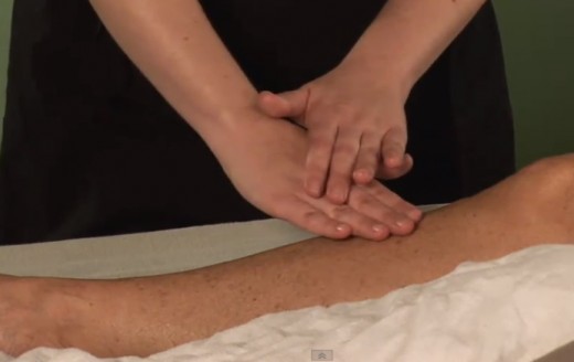
Wrapping the limb with a bandage can encourage the fluid to flow back towards the trunk of the body. Massage, pneumatic compression, and compression garments are other ways, which can be used to help in fluid movement. If the lymphedema is severe, the doctor may consider use of surgery procedure to remove the excess tissue in leg and arm.


However, this only helps in reducing severe pain and it cannot cure lymphedema. A complete decongestive therapy involving several of treatments like message, pneumatic compression, and compression garment may be recommended in managing the inflammation but this therapy is not suitable for people with high blood pressure, heart failure, paralysis, and blood clots.


Lymphedema Pictures


HIV Rash

HIV Rash
HIV is abbreviation of Human Immunodeficiency Virus which is a preceding condition of AIDS or Acquired Immunodeficiency Syndrome. This is a life threatening physical condition which destroys significant cells of the body.  The condition causes several symptoms. One of the common symptoms of the condition is HIV rash. This is a kind of rash on the skin which is commonly recorded in HIV patients. It is studied that around 80% of the HIV sufferers experience HIV rash symptoms. The rashes are often reddish in color and can be extremely itchy in nature. Appearance of these rashes may differ according to the severity of the disease. In most cases, raised small bumps are also observed.


HIV Rash Types


There is no concrete description of HIV rashes in the medical records. But HIV rashes can be divided into four classes which are as follows:


  • HIV rashes due to medicinal reaction:


There are antiretroviral medications that are known to cause skin rashes as side effects in some situations. These drugs are suggested as treatment for HIV infection and its associated symptoms. Some of such antiretroviral medicines include nevirapine, abacavir and amprenavir. These are few of the drugs that can lead to HIV rash. Ceasing the use of these medicines may possible ebb away rashes, but one should do this only after consulting your doctor.


  • HIV Rash of Acute nature


These are acute rashes which occur due to HIV infection. These rashes are noticed within few days from the time of affliction of the infection. There are other symptoms as well which are recorded along with these rashes. Chest, back, palms, etc, are the common regions for these rashes to occur; nevertheless, they can be seen on any part of the body. These kinds of rashes are often difficult to be diagnosed and may need thorough investigation through several tests.


  • Symptomatic HIV Rashes


The third phase of HIV infection may causes dermatitis which in turn leads to symptoms such as skin anomalies. These sort of rashes are usually pinkish or reddish in appearance as well as they may be itchy at times. These may last for 1 to 3 years according to records and estimation. Some of the common body regions where these symptomatic rashes can be found are groin, underarms, torso, facial skin and back.


  • HIV rashes due to Herpes


HIV sufferer may also have herpes and such individuals are prone to skin problems. If HIV is also accompanied by herpes then it becomes difficult for the patient to handle the condition. HIV and Herpes are STDs or Sexually Transmitted Disease. This makes the mortality rate extremely high. There are other symptoms of herpes which may be experienced along with these rashes. In such a situation the rashes are often filled with fluid. After few days these bumps bursts and spread further. The condition is very serious and if not controlled earlier, it may also lead to death.


Causes


HIV rashes mostly occur in people suffering from HIV infection. The HIV virus is a dreadful virus which can affect the tissues in the vaginal and anal lining. The virus can also affect mouth or eyes. There are three stages through which the infection progresses gradually. Occurrence of painful rashes as well as other flu-like symptoms indicates the primary stage of infection. The dermatologic symptoms may take place during seroconversion when noticeable antibodies are generated in oppose to infectious agents present in blood.


In some situations some antiretroviral drugs as mentioned above can also trigger allergic reaction or side effects causing rashes to appear. The Human Immunodeficiency Virus can severely influence and deteriorate the immune system causing the body to lose its ability to fight minor and major invaders of the body and infection. This also contributes to cause rashes on the skin.


Diagnosis


Chorionic skin rashes are often related with several skin issues which are usually difficult to diagnose through conventional physical screenings. In order to rule out HIV as underlying cause of rashes there are two routine tests suggested to the patient. The first test is specific to count of CD4 cells. People who are infected with the condition often have low CD4 count. The second test suggested is determining the amount of viral load in blood. Infected individual with high viral load are susceptible to reduced CD4 cells and faster progression of the condition as compared to those with low viral load. The efficacy of new treatments and medications can be evaluated by understanding the count of virus in blood.


Symptoms


HIV rashes has some features which makes it possible to recognized them; here are some aspects of HIV rashes


  • The patient suffering from the condition develops brown or else reddish lumps such as pimples. These appear similar to eczema and are in clusters

  • HIV infection may also lead to lesions due to skin peeling

  • In some cases HIV rashes can be itchy

  • HIV rashes can occur in almost any region of the body. In seldom cases even genitals, eyes as well as mouth is also infected

  • These HIV rashes may persist for up to 2 weeks but they are not transmissible


Other symptoms that may occur along with these rashes can hint that the skin condition is due to HIV.


  • Memory loss

  • Loose motions

  • Unexpected unknown fever

  • Pain in muscle

  • Body ache and cramps

  • Gland enlargement

  • Blurred vision

  • Lose of appetite and reduction in weight

  • Pain in joints


Treatment


HIV rash often resolves in around 2 weeks. However they may become dormant or asymptomatic. There is no confirmed cure for HIV infection but there are some hypotheses which can be a potential treatment in future. The treatment of HIV rash would depend on the cause of the condition. If it occurs due to medicinal side effects then ceasing the use of the medicine can help.  There are some non prescription medicines such as hydrocortisone cream which may alleviate the itchiness. Best treatment to deal with HIV rash can be recommended by your doctor alone. HIV is a serious health condition and adopting any measure to deal with it or its symptoms should be done only after approved by your doctor.


HIV Rash Pictures


Down Syndrome

Down Syndrome
Down syndrome is a genetic disorder, caused due to abnormality in the cell division involving chromosome 21. The disorder causes a permanent developmental delay, mental retardation and many other problems. The severities of Down syndrome vary and as such the problems of developmental delays also vary from mild to serious conditions. Learning difficulties are the common disability in Down syndrome.


When one of the three types of abnormal cell division involving chromosome 21 occur, Down syndrome is caused. The extra genetic material from chromosome 21, which is responsible for vital characteristic features of developmental difficulties are caused by all the three cell division abnormalities.


Early intervention and a better understanding of this disorder can help the children and adults affected by this syndrome.


Symptoms


The following are some of the signs and symptoms of Down syndrome:


  • Different and distinct facial features

  • A little/tiny head

  • Short neck

  • Flat face

  • Unusually upward slanting eyes that are different from the ethnic features.

  • Ears with a peculiar shape

  • A protruding tongue


Children with this disorder may also have the following additional signs:


  • Short, but broad hands along with a single crease in palm

  • Excessive flexibility

  • Poor muscle tone

  • Comparatively short fingers


Down syndrome affected children suffer from slow growth and are shorter in height. In comparison to other children, the Down syndrome affected children take double the time for growth and achieving developmental milestones such as crawling and sitting. They also elicit moderate mental retardation.


Children born with Down syndrome develop various complications as they grow. They are:


  • Leukemia

  • Obesity

  • Risk of dementia is more prevalent at an age of more than 40 along with higher rate of seizures.

  • Risk of infectious diseases due to weak immune system often causing pneumonia.

  • Heart disordersthat need surgery

  • Sleep apnea

  • Other problems like thyroid disorder, early menopause, gastrointestinal blockage, hearing loss, poor vision, skeletal disorder, seizures, and premature aging.


Causes


Generally, each cell of human being contains 23 pairs of chromosomes. The children get one chromosome of eachpair from their father and as well from their mother. The syndrome occurs when one of the three abnormal cell division connected to chromosome 21occurs. The cell division abnormalities result in additional genetic materialin chromosome 21 which is responsible for thedistinctive characteristics and developmental anomalies associated with the condition


The following are the three genetic variations that cause Down syndrome:


  • Mosaic Down syndrome:It is a rare form of syndrome that occurs whenever the children possess some cells with an extra copy of chromosome 21. The combination of normal cells with abnormal cells cause abnormal cell division after fertilization.

  • Trisomy 21:An infant with trisomy 21 possess three copies of chromosome 21, instead of two usual copies in all of his/her cells. This anomaly is caused due to abnormal cell division at the time of sperm cell or egg cell development. More than 90% of Down syndrome cases are caused by this trisomy 21.

  • Translocation Down syndrome:Whenever a part of the chromosome 21 remains attached to another chromosome (Translocated) at the time of conception or earlier, then it can cause Down syndrome. Children with translocation syndrome have additional material from the attached chromosome 21 in addition to the usual two copies of chromosome 21.The responsible factors for this syndrome are unknown.


Down syndrome is not inherited, but caused due to mistakes in cell division during the developmental stage of sperm or egg, or duringconception;except the translocation Down syndrome which may be passed on to the child from parent. Only 4% of the children are affected by translocation Down syndrome and only just half of them have inherited it from parents. The presence of some rearranged genetic material, even without extra genetic material, can also cause translocation Down syndrome. The chance of passing the translocation syndrome is 3% from father and 10 to 15% from mother.


An increased maternal age also increases the risk to conceiving a child with Down syndrome. The possibility expands along with the age. For example, the risk of conceiving a child with Down syndrome at the age of 35 is 1 in 400; while it is 1 in 35 at the age of 45.


Treatment


With the medical facility and treatment advancements, the average life expectancy of people with Down syndrome has increased up to 50 years.


Early intervention programs for children with Down syndrome will improve their abilities and their quality of life. The following are some of the treatments for Down syndrome affected children:


  • Administering of medicines as per the demands of the condition.

  • Early stimulation with appropriate sensory, motor, and other activitiesthat promote self-help.

  • Depending on the type of disorder, specialists from various fields will provide the needed treatments.

  • Self preparation to face the difficulties that may come while taking care of the affected child.

  • Consulting a genetic counselor before conceiving, if you are in the increased risk group

  • Seeking help from support groups


Down Syndrome life expectancy


People with Down syndrome have shorter than average life expectancy. However improvements in medical care and ongoing research have made it possible for people to have a longer life span. Back in 1912, the average life expectancy for Down Syndrome was 12 years; now it has increased to 60 years. It has been observed that most people who live up to their 40s and 50s, tend to get affected from neurodegenerative diseases like Alzheimer’s.


Pictures


Keratosis Pilaris

Keratosis Pilaris
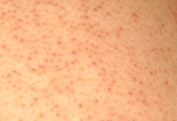
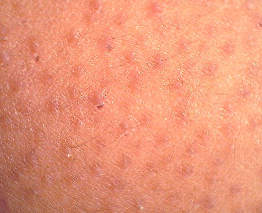
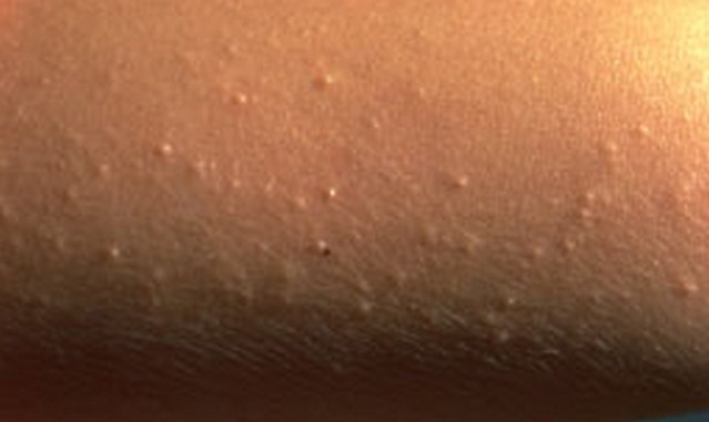
Keratosis Pilaris is a condition in which rough patches or blisters are noticed on the skin. These blisters are cluster of acne-line bumps. The condition is also referred to as ‘follicular keratosis’ or also as ‘chicken skin’. It is said to be an autosomal dominant medical condition. These dermatologic manifestations are often seen on arms, facial skin, thigh and other body regions. It is believed that blisters caused due to this condition do not appear on glabrous skin such as palm and sole of feet. In most cases the condition does not cause pain or itchiness. Majority of Keratosis pilaris cases are benign and ebb away with time without any treatment. Nevertheless, it is essential to consult your doctor as blisters and patches are also symptoms of serious medical issues.


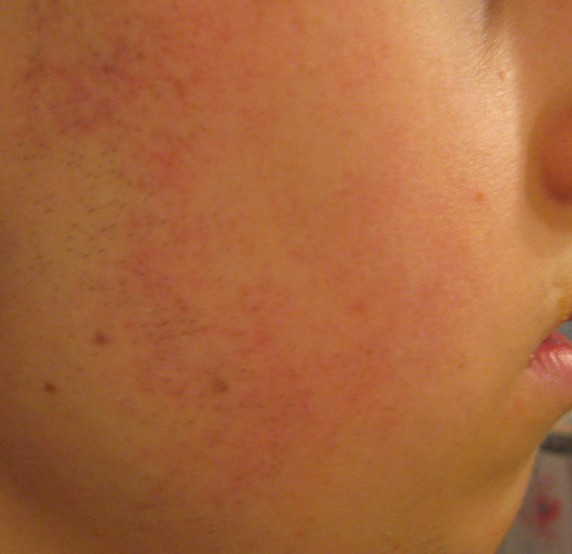
Keratosis pilaris when affects the face, it is often mistaken with conventional acne. Follicular keratosis is quite common; of the entire population approximately 40% to 50% people are affected with this dermatologic issue. It is more common in adolescent as there is 50% to 80% of the adolescent population suffering from the condition. Though the anomaly can affect both the genders, it is more common in women than men.


Types


There are different types of pilaris that may affect people. When red rashes appear on the facial region, particularly on cheeks, then it is possibly ‘Keratosis Pilaris Faceii’. Another type, in which red and inflamed elevated skin regions are seen on arms, legs, etc, is known as ‘Keratosis Pilaris Rubra’. ‘Keratosis Pilaris Alba’ is yet another kind in which rough bumpy skin is seen. However, in this condition there may be no irritation experienced.


These bumps are similar to goose-bumps in occurrence which is why most people may mistake Pilaris for these bumps. In fact goose-bumps are actually different as they result out of spontaneous muscle contraction. The only similarity in both of these conditions is that they occur on skin region where hair follicles are seen.


Symptoms


This condition is influential at any age however it is frequently diagnosed in children. In this condition there are numerous tiny boils occurring on the skin making the area rough. These bumps occur around the hair follicles on different body regions. Keratosis pilaris can also be inherited and may run in families. As mentioned above Keratosis Pilaris appears like goose-bumps or ‘chicken skin’. This condition is also seen in patients with atopic dermatitis and dry skin.  Here are some factors that will help you determine whether the condition is Keratosis Pilaris.


  • Tiny bumps appearing on body parts such as thighs, arms etc. These bumps may be reddish or white

  • Affected skin region would usually have dry and rough area

  • Blisters occurring on the affected area may cause itchiness

  • Keratosis specially exacerbates in winter which is also considered as a characteristics of the condition


When left untreated the condition may alleviate gradually without any medical intervention. However, it is better to consult a doctor and seek medical assistance as the condition may aggravate and become inflamed if left untreated, especially if occurring on face.


Causes 


It is discovered that Keratosis Pilaris is resulted due to keratin deposit. Keratin is a hard protein that ensures skin’s safety against infection as well as hazardous substances. Keratin develops into scaly plugs and at the opening of hair follicles causing obstruction of the follicle. There are many plugs that get developed under skin and appear as cluster of bumps on rough skin region. It is not yet understood what causes transformation of keratin into plugs. It is assumed that genetic influences along with possible skin irregularities may cause such a condition. Nevertheless, even healthy individuals may suffer from Keratosis Pilaris. The condition may aggravate if the skin is dry.


Is it necessary to see a doctor for Keratosis Pilaris?


This irregularity is not a serious medical anomaly usually. It often does not require any treatment. If the affected individual is concerned about his skin appearance then one can certainly consult a GP. It is better to directly approach a skin specialist technically known as ‘dermatologist’. The doctor may diagnose the condition usually by examining the skin physically and analyzing the scaly plugs.


Treatment


Unfortunately there is no treatment specifically decided for this condition. Doctors may often suggest self- care techniques and medicinal topical solutions to deal with the condition. The focus of the treatment remains on softening the keratin plug. Possible treatments for Keratosis Pilaris may include topical exfoliation. In this treatment there are topical creams suggested to the patient. These creams contain salicylic acid, alpha-hydroxy, urea moisturizer and lactic acid etc. Exfoliation helps in loosening and removal of dead skin cells. Topical retinoids are also suggested as it helps in preventing hair follicle obstruction due to plugs. In some cases repeated sessions of laser therapy may help in eradication of keratosis pilaris.


How to treat Keratosis pilaris with coconut oil?


The topical application of coconut oil has proved to be quite productive in treating this skin condition.  The results are visible in a matter of two  weeks or less.   Coconut oil alleviates the inflammation, and takes care of itching and redness, due to the presence of lauric acid that is known to be a natural moisturizer.  For greater effect, you can apply some coconut oil on the area affected by Keratosis pilaris, after you take a shower. Coconut oil body butter is also known to be increasingly beneficial to treat this condition. You can also apply the loofah dipped in coconut oil and moisturize the skin, during the bath.


Keratosis Pilaris Pictures


Excessive Sweating

Excessive Sweating Sweating is a normal process that helps in maintaining the right temperature for your body. Sweating occurs on the entire body but it would be more on the soles, palms and armpits and on the face. An individual is said to have excessive sweating if he/she sweats a lot irrespective of the temperature or activity level. In other words, a person will sweat excessively even if he is sitting idle and watching television. This would cause discomfort and embarrassment while taking part in social activities. In medical terms excessive sweating is called as hyperhidrosis and this can be managed by taking antiperspirants or sometimes by surgically removing the sweat glands from the body.


Types :


Excessive sweating can be classified into 3 types:-


Primary Hyperhidrosis :


Excessive sweating occurs in some body parts like palms, soles and armpits. There will be normal sweating in the rest of the body except few parts given above. This happens for unknown reason. Some believe that primary hyperhidrosis develop if there is an overactive sweat gland in these areas alone.


Secondary Hyperhidrosis :


This type of excessive sweating is rare. Sweating in excess will be there in particular part like hand or leg. Here there will be a definite cause for sweating.


Generalized Hyperhidrosis :


In this type excessive sweating will be there in all over the body. Again there will be underlying cause for hyperhidrosis.


Causes :


When a person sweats, the body senses excess of heat. The brain would automatically send signals to nervous system which would trigger secretion of sweat by sweat glands. Excessive sweating is present if the nervous system becomes overactive releasing excess of sweat. A person sweats more if he is stress or anxiety. It can happen due to genetic factor since the same gene runs in families. Intake of certain medications, underlying medical issues like overactive thyroid gland, heart problems, low blood sugar and infectious diseases can cause excessive sweat.


Symptoms :


There will be excess of sweat in the hands, feet and armpits and even on the face. For extreme cases, excess of sweat will soak the clothing and drip off from the hands. Excessive sweating does not cause any complications but in rare cases it can affect the skin condition causing infection and rashes.


Diagnosis :


Your doctor will collect the medical history, health condition and medication you are taking at present. In addition he may order for certain lab tests like blood and urine analysis. Special sweat test is done using powder that changes its color when there is excessive sweating. This is called as thermoregulatory test.


Treatment :


The method of treatment depends on the causative factor. It is good to try some home remedies and over the counter medications before going to your doctor. Your doctor would suggest high strength of antiperspirants which can be used before you go to bed. Initially it can cause skin irritation but you would feel better in the morning. If the nervous system is causing the problem your doctor may give pills to block the neurotransmitters. This would eventually stop the communication of the nervous system to the sweat glands. However the drugs can cause some adverse effect.


You can try using aluminum chloride antiperspirants like Anhydrol Forte or Driclor to control excessive sweating. Botox injection is used to block the nerves to stop the function of sweat glands. But it is expensive and the result would not be long lasting. Surgery is done for severe cases of hyperhidrosis. In a process called iontophoresis the doctor would send electric current (in low dose) on the hands, feet and armpit keeping that part in the water. But this therapy has to be repeated for weeks and months to get relief from excessive sweating.


In some cases, the surgeon would remove the sweat glands from particular part of the body like armpits or hands. This process is simple and can be done through minor incisions. In rare cases nerve surgery is done to provide clamps on the spinal nerves to block the signals.


Home Remedies :


First you should check if the soaps you are using are strong since it can cause skin irritation. Hence you can replace it with mild soap. Avoid eating hot and spicy foods since it can trigger excess sweat. Apply antiperspirant daily on the armpits, hands and legs where there is excess of sweat.


Change clothes at least twice a day if you are sweating a lot. Avoid wearing tight clothing. You can use dress shields to absorb sweat easily. Change socks regularly and apply absorbent powder liberally on your feet. Choose right size of shoes that give space for breathing.


Ganglion Cyst Wrist

Ganglion Cyst Wrist Small lumps that develop on the tendons of your hands and wrist are called ganglion cyst wrist. Apart from wrist area they can develop on the ankles and feet also. They are tiny round shaped lesions filled with pus like fluid. The appearance of lumps on the wrist area can sometimes interfere with the normal joint movement. Ganglion cyst on the wrist can be aspirated using a needle to drain off the fluid inside. But for many people, the cyst would resolve after some days without requiring any treatment. Ganglion cyst largely occurs in women for no apparent reason. It either occurs as single or in groups covering a tendon or joint.


Symptoms :


Ganglion cysts are tiny fluid filled blisters that appear on the tendons of your hands. For some people they can also develop on the feet and ankles. The size of the cyst is small measuring half inch or one inch in diameter. For some people the size of the cyst is so tiny that it can be felt only if the area is pressed. Ganglion cyst sometimes can be painful and for some people it can cause tingling sensation or numbness on the affected wrist.


Causes :


Exact cause of formation of ganglion cyst wrist is not known. It appears from a joint area or tendon of the wrist to grow like a lesion or lump on the hand.


Who are at risk? :


Ganglion cyst wrist can develop in any age but it is more likely to occur on middle aged women than children. Individuals with arthritis problems are prone to develop this cyst on their joints. People with tendon injury have increased chance for developing ganglion cyst.


Test :


Your doctor will complete the physical exam of the cyst on your wrist or feet. He would gently apply pressure on the area to check if you have pain.


Pictures of Ganglion Cyst Wrist :


Images, Pics, Pictures and Photos of Ganglion Cyst Wrist


Ganglion Cyst Wrist Ganglion Cyst Wrist Ganglion Cyst Wrist Ganglion Cyst Wrist Ganglion Cyst Wrist Ganglion Cyst Wrist


Treatment :


Ganglion cyst wrist does not require any treatment in most of the cases, since it will disappear within few days. Over the counter drugs like Ibuprofen can be used if there is pain. Aspiration is the procedure used for treating cyst. Your doctor would insert a needle directly on the cyst to drain out the liquid from the cyst. He would then give a steroid injection on the area to facilitate healing and to prevent further cyst formation.


About 75% of the people are getting relieved from the cyst using aspiration procedure. In rare cases the cyst will recur after some months. Surgery is done for removing the cyst only if the person suffers from pain or numbness in the affected area. Arthroscopic procedure is largely used for surgical removal of the ganglion cyst. After the process, you need to wear a splinter or brace so that there is no mobility on that area. Once the cyst is removed by aspiration or surgery the pain will gradually reduce since the nerve will not feel any pressure anymore.