Epidermoid Cyst – Symptoms, Causes, Treatment, Removal, Pictures
These are bumps that are small and develop on face, trunk, and neck. They grow very slowly and are normally painless. These are often referred to as sebaceous cysts. But real sebaceous cysts are rarer than these cysts.
The majorities of these cysts do not create any problems nor need any treatment. Often they are of concern cosmetically or they can rupture and become infected. This is when they normally are removed surgically.
These cysts are most of the time non-cancerous but there are those rare cases when they do turn into cancers of the skin. Since this happens so rarely, epidermoid cysts normally are not biopsied except when they have characteristics that are unusual that can advocate a problem that is more serious.
Epidermoid cysts:
Often these cysts have an opening in the center – which is the vestige of a follicle of hair from where these cysts originate from – that is clogged by a tiny black head. An individual is often able to squash out a very thick and cheesy matter thru the opening but due to the risk of an infection as well as scarring it really is best to see a skin specialist.
Milia
These are deep-seated, tiny whiteheads that seem to never move to the skin surface – are actually epidermoid cysts but miniature. They are very common in elderly ladies and in those males who have substantial damage due to the sun on temples as well as cheeks. They may also be aggravated or caused by long time use of cosmetics or creams that are oil-based.
Symptoms and signs of infection, which may occasionally happen, consist of:
Often an individual can develop small bumps on the scalp that often looks like epidermoid cysts. These are in most cases trichilemmal or pilar cysts that normally have walls that are thicker than epidermoid cysts are and will in almost all cases move easily under the skin. The linings of these cysts also differ to some extent from an epidermoid cyst.
Although most epidermoid cysts are not dangerous, an individual may still want them removed for cosmetic motivation. A person should see their doctor if the cyst:
The skin surface – also known as this epidermis – consists of very thin, protecting cell layer which the body sheds continuously. Most of these epidermoid cysts develop when these cells on the surface rather than exfoliating normally, instead move deep into the skin and continue to multiply. More often, this happens in the areas having very small follicles of hair and much bigger oil glands or sebaceous glands – on the groin area, upper back, neck and face.
These cells of the epidermal form the cysts walls and then begin to secrete keratin, a protein, in the interior. This keratin is yellowish, thick matter than often drains from the cyst.
Factors that lead to this atypical production of cells include:
Damage to hair follicle
Every hair develops from a hair follicle which is a slight pocket of skin of the dermis which is modified and is the skin layer underneath the epidermis. These follicles become injured by damage from surgical wounds, or abrasions and can then become clogged by cells of the surface.
A sebaceous gland that is ruptured
Located just over the follicles of hair, sebaceous glands create sebum which is the oil lubricating the skin and coating every hair shaft. These glands can rupture very easily by skin conditions that are inflammatory, particularly acne, making this site likely for epidermoid cysts to develop.
Heredity
Epidermoid cysts may develop in individuals with Gardner’s syndrome – an uncommon inherited condition that causes developments in the colon, or basal cell nevus syndrome which is a genetic disease leading to some serious defects.
Nearly any individual can have one or even more epidermoid cysts, but there are some factors which can make an individual more prone for development:
Past puberty
They will occur at any age, epidermoid cysts are very rare before puberty
http://www.Symptoms-Causes-treatment.blogspot.com detect diseases at an early stage symptoms, and find out the causes and treatments best suited.
Being a male
Men are much more prone to have these cysts.
History of acne
These cysts are very common in individuals who have had acne
Significant sun exposure
Milia normally occur in those men and women with a long past of sun exposure.
Skin injuries
Any crushing or traumatic injury to the skin such as slamming the hand in a car door can increase the risk of developing epidermoid cyst.
There are rare cases of epidermoid cysts that give rise to squamous and basal cell cancers of the skin. Since this occurs so rarely, these cysts normally are not biopsied unless they become solid, immobile, infected or develop other unusual characteristics that can suggest a problem that is more serious. Aside from cancer, other complications can include:
Inflammation
These cysts may become swollen and tender, even when they are not infected. Cysts that are inflamed are hard to remove and the physician normally will postpone any treatment until the inflammation diminishes.
Rupture
A cyst that ruptures can lead to boil-like abscesses that need quick treatment.
Infection
These cysts may become infected naturally or after a rupture.
Those cysts that do not cause functional or cosmetic problems are normally not treated. When a cyst is ruptured, infected or inflamed, these options for treatment occur:
Injections corticosteroid
The primary care physician or skin specialist can inject any epidermoid cyst that is inflamed but not infected, with a corticosteroid to aid in reducing any inflammation.
Incision and drainage
With this procedure, the physician makes a tiny incision in the cyst and expresses any contents. Even with this procedure being very easy and quick, cysts can often recur even later after this treatment.
There are several options for removal of these cysts. They include:
Total excision
This technique is surgical and gets rid of the entire cyst. This prevents any return. Excision is more effective if the cyst is not inflamed. The physician will usually first treat any inflammation with steroids antibiotics or incision with drainage before performing excision. There will usually be a waiting of 4 to 6 weeks after the inflammation resolves. Total excision will need sutures. The physician will remove any sutures in the face in 7 days or so of the procedure and will take out the sutures in other areas of the body within 1 to 2 weeks.
Minimal excision
Many physicians prefer this option because it not only removes the entire cyst wall but also causes very negligible scarring. The physician will make a small incision in the cyst, expresses any substances, and then eliminates the wall of the cyst thru the incision. This minor wound is normally allowed to heal naturally.
Lasers
In order to minimize any scarring, the physician can use carbon dioxide laser to evaporate the cyst on the face or additional areas that are sensitive.

Epidermoid cyst on chin
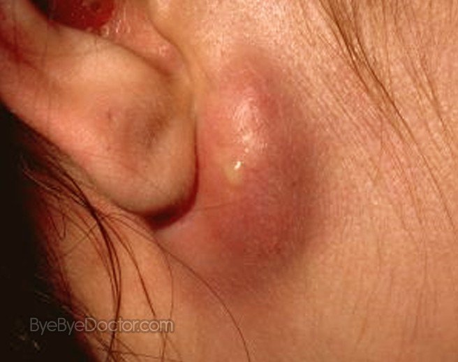
 near ear
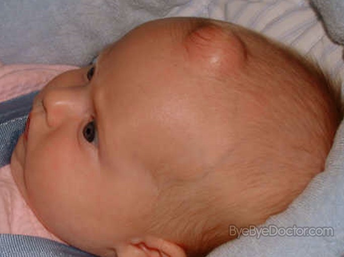
 in babies
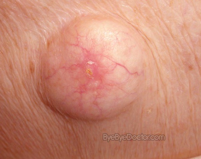
 on skin
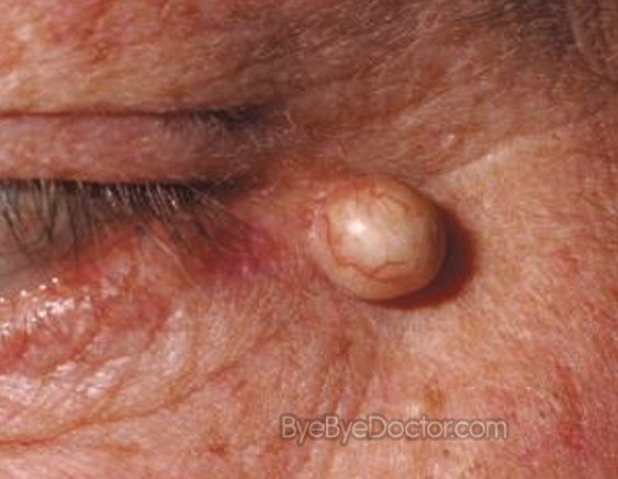
 near eye
What is an Epidermoid Cyst?
These are bumps that are small and develop on face, trunk, and neck. They grow very slowly and are normally painless. These are often referred to as sebaceous cysts. But real sebaceous cysts are rarer than these cysts.
The majorities of these cysts do not create any problems nor need any treatment. Often they are of concern cosmetically or they can rupture and become infected. This is when they normally are removed surgically.
These cysts are most of the time non-cancerous but there are those rare cases when they do turn into cancers of the skin. Since this happens so rarely, epidermoid cysts normally are not biopsied except when they have characteristics that are unusual that can advocate a problem that is more serious.
Epidermoid Cyst Symptoms
Epidermoid cysts:
- Normally yellow or white, but some individuals with skin that is darker can have cysts that are pigmented
- Are round, small bumps or cysts which are easily moved with the fingers
- Range in sizes from smaller than ¼ of an inch to up to 2 inches in diameter
- Develop on almost all parts of the body, including the fingernails however are mostly developed on the neck, trunk or face.
Often these cysts have an opening in the center – which is the vestige of a follicle of hair from where these cysts originate from – that is clogged by a tiny black head. An individual is often able to squash out a very thick and cheesy matter thru the opening but due to the risk of an infection as well as scarring it really is best to see a skin specialist.
Milia
These are deep-seated, tiny whiteheads that seem to never move to the skin surface – are actually epidermoid cysts but miniature. They are very common in elderly ladies and in those males who have substantial damage due to the sun on temples as well as cheeks. They may also be aggravated or caused by long time use of cosmetics or creams that are oil-based.
Symptoms and signs of infection, which may occasionally happen, consist of:
- Redness, tenderness and swelling surround the cyst
- Thick, yellow matter which drains from the cyst that in many cases has a bad odor
A condition that is similar looking
Often an individual can develop small bumps on the scalp that often looks like epidermoid cysts. These are in most cases trichilemmal or pilar cysts that normally have walls that are thicker than epidermoid cysts are and will in almost all cases move easily under the skin. The linings of these cysts also differ to some extent from an epidermoid cyst.
Although most epidermoid cysts are not dangerous, an individual may still want them removed for cosmetic motivation. A person should see their doctor if the cyst:
- Rapidly grows
- Becomes painful
- Ruptures
- Develops in an area where it is constantly irritated
Epidermoid Cyst Causes
The skin surface – also known as this epidermis – consists of very thin, protecting cell layer which the body sheds continuously. Most of these epidermoid cysts develop when these cells on the surface rather than exfoliating normally, instead move deep into the skin and continue to multiply. More often, this happens in the areas having very small follicles of hair and much bigger oil glands or sebaceous glands – on the groin area, upper back, neck and face.
These cells of the epidermal form the cysts walls and then begin to secrete keratin, a protein, in the interior. This keratin is yellowish, thick matter than often drains from the cyst.
Factors that lead to this atypical production of cells include:
Damage to hair follicle
Every hair develops from a hair follicle which is a slight pocket of skin of the dermis which is modified and is the skin layer underneath the epidermis. These follicles become injured by damage from surgical wounds, or abrasions and can then become clogged by cells of the surface.
A sebaceous gland that is ruptured
Located just over the follicles of hair, sebaceous glands create sebum which is the oil lubricating the skin and coating every hair shaft. These glands can rupture very easily by skin conditions that are inflammatory, particularly acne, making this site likely for epidermoid cysts to develop.
Heredity
Epidermoid cysts may develop in individuals with Gardner’s syndrome – an uncommon inherited condition that causes developments in the colon, or basal cell nevus syndrome which is a genetic disease leading to some serious defects.
Nearly any individual can have one or even more epidermoid cysts, but there are some factors which can make an individual more prone for development:
Past puberty
They will occur at any age, epidermoid cysts are very rare before puberty
http://www.Symptoms-Causes-treatment.blogspot.com detect diseases at an early stage symptoms, and find out the causes and treatments best suited.
Being a male
Men are much more prone to have these cysts.
History of acne
These cysts are very common in individuals who have had acne
Significant sun exposure
Milia normally occur in those men and women with a long past of sun exposure.
Skin injuries
Any crushing or traumatic injury to the skin such as slamming the hand in a car door can increase the risk of developing epidermoid cyst.
There are rare cases of epidermoid cysts that give rise to squamous and basal cell cancers of the skin. Since this occurs so rarely, these cysts normally are not biopsied unless they become solid, immobile, infected or develop other unusual characteristics that can suggest a problem that is more serious. Aside from cancer, other complications can include:
Inflammation
These cysts may become swollen and tender, even when they are not infected. Cysts that are inflamed are hard to remove and the physician normally will postpone any treatment until the inflammation diminishes.
Rupture
A cyst that ruptures can lead to boil-like abscesses that need quick treatment.
Infection
These cysts may become infected naturally or after a rupture.
Epidermoid Cyst Treatment
Those cysts that do not cause functional or cosmetic problems are normally not treated. When a cyst is ruptured, infected or inflamed, these options for treatment occur:
Injections corticosteroid
The primary care physician or skin specialist can inject any epidermoid cyst that is inflamed but not infected, with a corticosteroid to aid in reducing any inflammation.
Incision and drainage
With this procedure, the physician makes a tiny incision in the cyst and expresses any contents. Even with this procedure being very easy and quick, cysts can often recur even later after this treatment.
Removal
There are several options for removal of these cysts. They include:
Total excision
This technique is surgical and gets rid of the entire cyst. This prevents any return. Excision is more effective if the cyst is not inflamed. The physician will usually first treat any inflammation with steroids antibiotics or incision with drainage before performing excision. There will usually be a waiting of 4 to 6 weeks after the inflammation resolves. Total excision will need sutures. The physician will remove any sutures in the face in 7 days or so of the procedure and will take out the sutures in other areas of the body within 1 to 2 weeks.
Minimal excision
Many physicians prefer this option because it not only removes the entire cyst wall but also causes very negligible scarring. The physician will make a small incision in the cyst, expresses any substances, and then eliminates the wall of the cyst thru the incision. This minor wound is normally allowed to heal naturally.
Lasers
In order to minimize any scarring, the physician can use carbon dioxide laser to evaporate the cyst on the face or additional areas that are sensitive.
Epidermoid Cyst Pictures

Epidermoid cyst on chin
 near ear
 in babies
 on skin
 near eye
No comments:
Post a Comment